Write to us
the fields marked with * are mandatory

Narrowed or damaged blood vessels often remain unnoticed for a long time – until serious complications occur. Early and reliable diagnostics that consider the entire body are therefore particularly important. Modern radiological procedures now make it possible to visualize vascular changes precisely. Anyone looking for precise and gentle diagnostics can find an important basis for further medical care with whole-body MR angiography at our private radiology practice in Düsseldorf.
Whole-body check of the blood vessels
Visualizes arteries and veins from head to toe
Early warning system for your blood vessels
Detects vascular narrowing, aneurysms, thromboses and malformations early and reliably
Ideal for unexplained symptoms
Helps identify the cause of symptoms such as dizziness, leg pain or high blood pressure
Without surgery
The blood vessels are visualized painlessly and non-invasively
Gentle imaging with fewer risks
No catheter-related complications such as bleeding or embolism and a lower risk of side effects

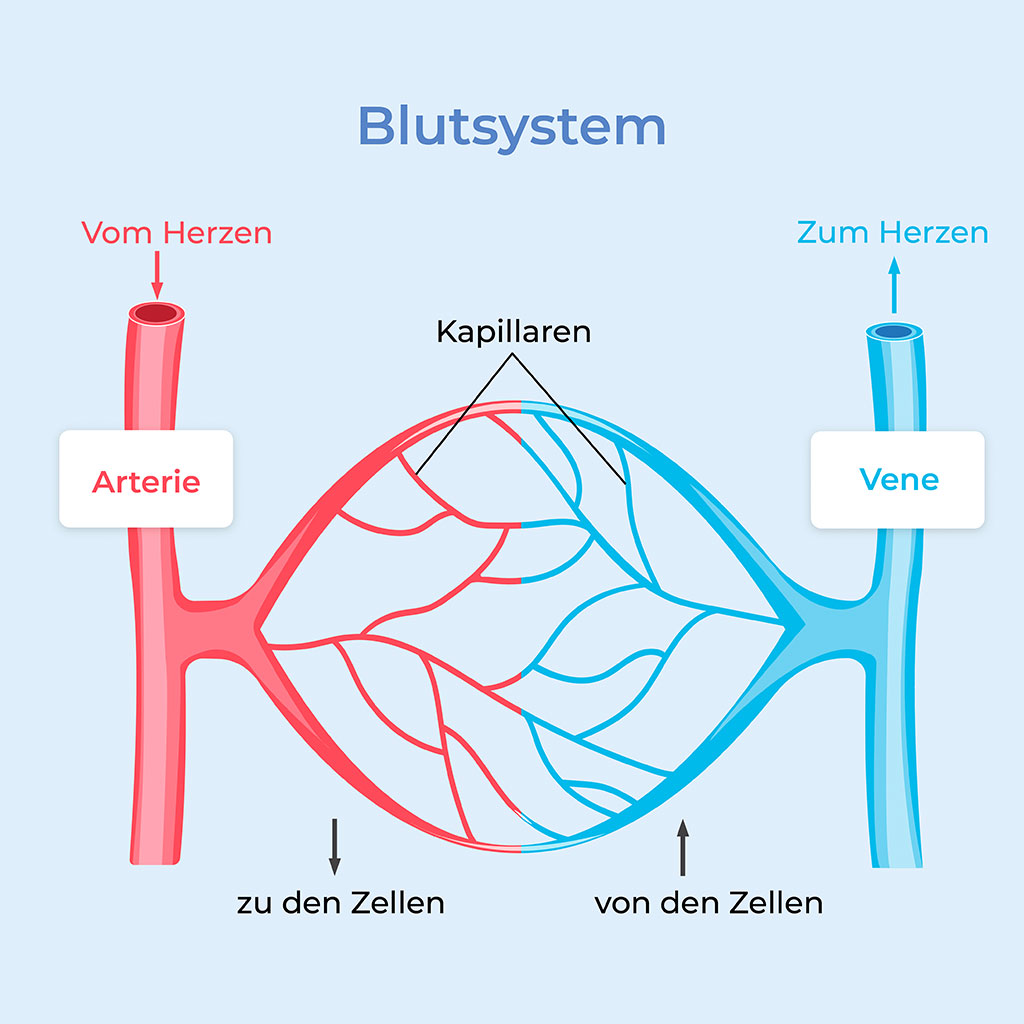
The human body has three main types of blood vessels, each with specific functions: arteries, veins and capillaries.
They transport oxygen-rich blood from the heart to the different regions of the body. The most important arteries of particular medical relevance include:
The coronary arteries play a central role in the function of the heart. They branch off from the main artery (aorta) directly above the aortic valve and run around the heart like a crown. Their function is to continuously supply the heart muscle with oxygen-rich blood and nutrients.
Depending on their anatomical course, several major branches of the coronary arteries are distinguished, including:
The carotid arteries are important blood vessels that supply the brain. They run along both sides of the neck and transport oxygen-rich blood from the heart toward the head. Their main function is to supply the brain, eyes and parts of the face with vital oxygen and nutrients.
The carotid arteries include the common carotid artery (Arteria carotis communis), which divides into two branches at the level of the larynx:
As the largest blood vessel in the human body, the aorta is of central importance for circulation throughout the body. It originates directly from the left ventricle and transports oxygen-rich blood under high pressure into the systemic circulation. Through numerous branches, it distributes blood to all organs and tissues to maintain their function.
Anatomically, the aorta is divided into several sections:
The arteries of the legs supply the muscles and tissues of the lower extremities with oxygen-rich blood. They carry blood from the abdominal aorta through the pelvic region and down to the feet, thereby ensuring blood flow to the legs – both at rest and during physical activity.
The most important sections of the leg arteries include:
The human body has an extensively branched venous system that transports oxygen-poor blood from the body back to the heart. The venous system includes:
It collects venous blood from the head, neck, upper extremities and chest and carries it to the right atrium of the heart.
Important tributaries include:
This vein transports blood from the abdomen, pelvis and legs back to the heart.
The tributaries of the inferior vena cava include:
They return oxygen-poor blood from the legs to the heart. They consist of superficial, deep and connecting (perforating) venous systems.
At the center of this system is the hepatic portal vein (Vena portae hepatis), which is formed by several large abdominal veins. Unlike other veins, it does not carry blood directly to the heart, but first to the liver. Only after passing through the liver does the filtered blood enter the inferior vena cava via the hepatic veins and return to the regular circulation.
This is a specialized, expanded venous system within the dura mater that does not have the typical structure of a vein. Instead, it consists of tube-like blood channels running between the two layers of the dura mater and plays a central role in draining blood from the skull.
The cerebral veins collect oxygen-poor blood from the different regions of the brain and carry it into the venous sinuses.
The most important include:
These smallest and finest blood vessels in the human body form a dense network between arteries and veins. Capillaries run through almost every tissue.
Their main function is the exchange of substances: oxygen and nutrients pass from the blood into the surrounding tissue, while carbon dioxide and waste products from the cells are absorbed into the capillaries and transported away. The walls of capillaries consist of a single layer of cells, which facilitates the rapid exchange of substances.
Properly functioning capillaries are essential for healthy blood circulation and maintaining organ function.
If blood vessels can no longer perform their functions properly, this may have serious health consequences:
Whole-body angiography is a modern imaging procedure that allows detailed visualization of blood vessels throughout the body. The aim of the examination is to detect harmful vascular changes at an early stage – ideally before symptoms occur. It is suitable both for preventive examinations in patients with risk factors and for the targeted investigation of existing symptoms.
At our private radiology practice in Düsseldorf, we use whole-body angiography with MRI (magnetic resonance imaging) to visualize the blood vessels throughout the body precisely and gently. This method does not use X-rays and is therefore particularly well tolerated. The examination is non-invasive and painless and provides high-resolution images of all relevant vascular regions, enabling a meaningful medical assessment. Based on these findings, your treating physician can plan the most appropriate treatment for you.
Whole-body MR angiography offers a safe way to diagnose vascular diseases. At our private radiology practice in Düsseldorf, you benefit from state-of-the-art technology, high diagnostic precision and personal, reassuring care.
Before the examination begins, we have a detailed consultation with you. We discuss your medical history, existing risk factors and any pre-existing conditions. We also provide comprehensive information about the procedure and the contrast agent used.
You remove all metallic objects because MRI uses a strong magnetic field. As a rule, no special preparation is necessary. If a contrast agent is used, it is administered beforehand via a vein in your arm.
During the examination, you lie comfortably on an examination table that is slowly moved into the MRI scanner. Whole-body angiography is performed in several sections so that all relevant vascular regions can be visualized in detail. We gradually image the blood vessels in the neck, coronary vessels, aorta, renal and pelvic vessels as well as the arteries and veins of the legs.
Immediately after the examination, you can leave the examination room and get dressed again. Our radiologists then analyze the imaging data with the utmost care. We prepare the report promptly and make it available both to you and to your treating physician.
Whole-body MR angiography is not only an important diagnostic procedure for existing symptoms – it is also particularly suitable for preventive healthcare.
Particularly in people with risk factors such as high blood pressure, diabetes, elevated blood lipid levels, a history of smoking or a family history of vascular disease, early vascular diagnostics can be important for health. Whole-body angiography can also provide valuable guidance in patients with non-specific symptoms such as dizziness, chest pain or calf pain when walking.
The aim of the examination is to detect pathological changes in the blood vessels at an early stage before serious complications such as a heart attack or stroke occur.
Narrowing of blood vessels is frequently caused by deposits known as plaques, which form in the vessel walls as a result of atherosclerosis.
Depending on where these narrowings occur, they can have very different and sometimes serious consequences. If, for example, a carotid artery becomes narrowed (carotid stenosis), the risk of a stroke increases significantly. If the coronary arteries are affected (coronary stenosis), angina pectoris or even a heart attack may occur. Narrowing of the leg arteries frequently causes peripheral arterial disease, in which those affected have to stop regularly when walking because of pain.
The renal arteries can also be affected – stenosis in this area can cause high blood pressure or significantly worsen existing hypertension.
Aneurysms are localized bulges in the vessel wall that occur particularly frequently in the aorta – the main artery in the abdominal or chest area. Such vascular changes can also occur in the brain.
The danger of aneurysms is that they often cause no symptoms for years and therefore remain unnoticed. However, if an aneurysm ruptures, this can lead to life-threatening internal bleeding. Abdominal aortic aneurysms, thoracic aortic aneurysms and cerebral aneurysms are among the common forms.
A complete vascular occlusion is frequently caused by a blood clot (thrombus) that suddenly blocks the flow of blood.
The consequences can be serious: if an acute occlusion occurs in an artery of the leg, ischemia may develop, meaning that the tissue receives an insufficient supply of oxygen – a medical emergency. If a thrombus blocks an artery in the brain, it may cause a stroke. A pulmonary embolism is also particularly dangerous when a clot breaks away from a deep vein in the leg and travels to the lungs.
Inflammation of the vessel walls can, for example, lead to narrowing or direct damage to the affected blood vessels.
Such inflammatory processes do not only affect individual regions of the body, but can also have systemic effects. Takayasu arteritis, for example, is associated with chronic inflammation of large arteries, while giant cell arteritis mainly affects older people and increases the risk of serious complications such as visual disturbances or stroke.
ANCA-associated vasculitis can also affect the vascular system and lead to a wide range of often multisystemic symptoms.
The venous system can also be reliably visualized using whole-body angiography and can provide important information about possible diseases.
For example, it can be used to detect deep vein thrombosis (DVT), in which a blood clot forms in the deep veins of the legs – a potentially dangerous situation because the clot can travel to the lungs and cause an embolism.
In chronic venous insufficiency, occlusions and collateral circulation can also be identified. These are alternative pathways formed by the body to compensate for impaired blood flow.
Precise visualization of these changes is important for targeted treatment and for preventing further complications.
Whole-body angiography at our radiology practice in Düsseldorf can also visualize congenital vascular malformations as well as malformations acquired during a person’s lifetime.
These include, for example, arteriovenous malformations (AVMs) – direct connections between arteries and veins in which the capillary network that would normally lie between them is absent. These can significantly alter blood flow and cause symptoms such as pain, bleeding or neurological deficits.
Venous malformations can also be detected. These are abnormal developments of the veins and frequently cause swelling, a feeling of pressure or visible skin changes.
MR angiography is also particularly suitable for monitoring vascular procedures that have already been performed. It allows us, for example, to reliably check whether stents or bypass vessels remain open and functional.
The position and condition of vascular prostheses can also be assessed precisely with the examination – without radiation exposure. In addition, the examination provides important information for planning further interventions, for example if another vascular treatment becomes necessary. In this way, angiography makes an important contribution to safe and comprehensive follow-up.
As you can see, whole-body angiography makes it possible to detect a wide range of diseases at an early stage, precisely assess their extent and make well-founded treatment decisions.
Simply contact us to arrange an appointment for whole-body angiography at our practice in Düsseldorf!

the fields marked with * are mandatory
For patients with public health insurance
For patients with private insurance and those paying out of pocket