Write to us
the fields marked with * are mandatory

Not every change in breathing can be clearly identified with basic examinations. If symptoms persist or findings do not provide a clear picture, conventional procedures can reach their limits. In these situations, modern cross-sectional imaging offers important additional information. It enables differentiated visualization of the airways and can reveal changes that are not visible with conventional methods. Computed tomography provides reliable information in cases of unclear respiratory problems and creates a sound basis for further diagnostic or therapeutic decisions.
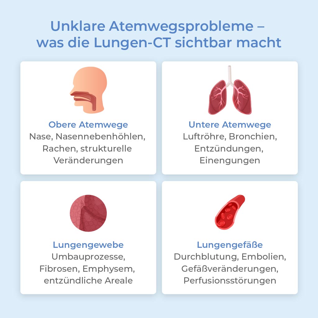
Breathing disorders cannot always be traced back to a single cause. Depending on which part of the airways is affected and whether neighboring structures are involved, different clinical conditions may be considered. Several groups of possible causes therefore need to be taken into account.
These include:
The upper airways include the nose, paranasal sinuses, pharynx and larynx. Inflammation of the sinuses, swelling of the mucous membranes, polyps or narrowing in the laryngeal area can significantly impair breathing. Anatomical features such as a deviated nasal septum, enlarged turbinates, congenital narrowing in the pharynx or structural changes in the larynx can also play a role. Scarring after previous surgery may be another possible cause.
The lower airways include the trachea, bronchi and lungs. Common causes of symptoms include chronic inflammation of the bronchi, bronchial asthma, chronic obstructive pulmonary disease or tumors. Acute infections, scarring or narrowing of the airways can also impair oxygen uptake.
Not all breathing problems originate directly in the bronchi. Structural changes in the lung tissue, for example due to fibrosis, emphysema or inflammatory changes, can restrict gas exchange. Such changes often remain unnoticed in their early stages.
The blood vessels of the lungs also influence breathing. Circulatory disorders, vascular changes or blood clots can cause shortness of breath even when the airways themselves appear unobstructed.
Some respiratory symptoms cannot be clearly classified even after initial examinations. The available findings do not adequately explain the symptoms or do not result in a consistent overall clinical picture.
Several factors often interact. Inflammatory processes, structural changes, circulatory disorders or abnormalities of the lung tissue can overlap and make classification more difficult. Some changes also develop gradually and remain undetected with standard procedures in their early stages.
When respiratory problems first occur, basic examinations provide important initial guidance. In general practice or pulmonology, the focus is on a detailed medical history, physical examination and listening to the lungs. Pulmonary function tests can provide additional information about narrowing of the airways or reduced lung performance.
Blood tests may indicate inflammation or infection. A chest X-ray is often used as the first imaging examination and can show larger changes in the lungs or heart region.
If the findings remain unremarkable or do not adequately explain the symptoms, more detailed diagnostic imaging may be recommended.
Computed tomography of the lungs, as offered at our radiological prevention center in Düsseldorf, complements examinations such as X-ray imaging and pulmonary function testing with details that are not visible using these standard procedures. While basic examinations mainly provide information about overall lung function, CT enables direct assessment of lung tissue, the bronchi and blood vessels.
The examination uses high-resolution cross-sectional images that can reveal even subtle structural changes. Areas of increased density, scarring, inflammatory processes or vascular abnormalities can be identified precisely and differentiated from one another. Changes that cause no clear symptoms in their early stages can also become visible.
This provides an important advantage in the diagnosis of unclear respiratory problems: lung CT not only confirms that symptoms are present, but can also provide information about what may be causing them. These findings can help guide the next steps.
Important to know: lung CT does not visualize the upper airways. Questions involving the nose, paranasal sinuses, pharynx and larynx require other procedures, such as CT of the paranasal sinuses or MRI of the neck / pharynx / larynx.

the fields marked with * are mandatory
At Radiology Düsseldorf, lung CT provides a high-resolution method for structural assessment of the lungs, bronchi and blood vessels. The examination supports reliable clarification of unclear respiratory symptoms and provides a sound basis for further medical assessment. Computed tomography can also be used for general prevention and early detection, for example of lung cancer.
Simply contact us to arrange an appointment!
Image material: © DimaBerlin / stock.adobe.com, Gaurav / stock.adobe.com