Write to us
the fields marked with * are mandatory

Many changes in the blood vessels develop over years. The body can compensate for minor restrictions in blood flow for a long time. Nevertheless, warning signs may indicate problems in the arteries. These include pressure in the chest during exertion, recurring dizziness or pain in the legs. Elevated cholesterol levels or a family history can also indicate that deposits may be forming in the vessels. Early assessment can help identify narrowing in good time. Modern radiological examinations visualize blood vessels and allow circulatory disorders and vascular changes to be assessed carefully. This can help identify risks of heart attack, stroke or circulatory problems in the legs at an early stage.
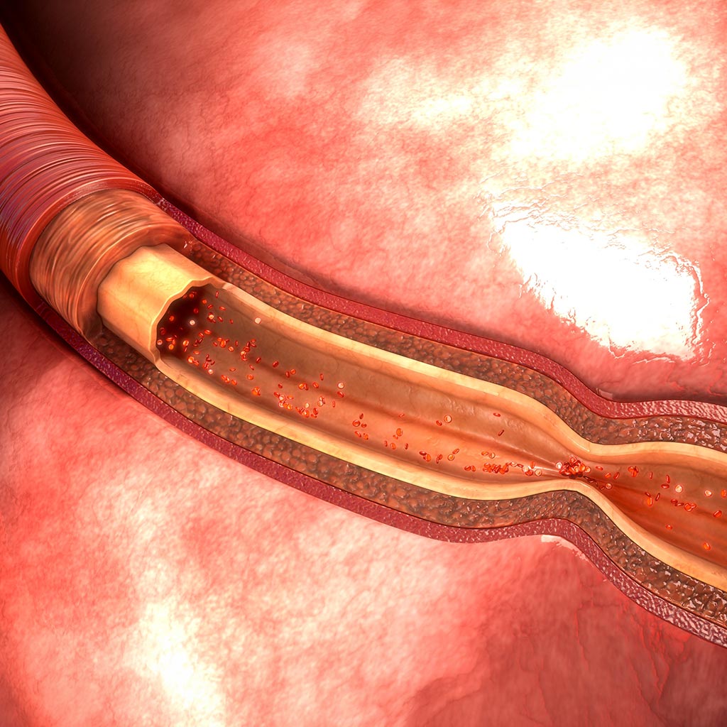
Arteries transport large amounts of blood through the body every day. The heart, brain, kidneys and muscles depend on blood flowing freely through these vessels. If the inner wall of an artery changes, however, the diameter of the vessel can gradually become smaller.
So-called plaques play a central role. These are deposits that form in the vessel wall. Plaques consist mainly of fatty substances, particularly cholesterol, as well as calcium, connective tissue and inflammatory cells. Over time, these components accumulate in the inner vascular layer. The vessel wall thickens, loses elasticity and increasingly protrudes into the inside of the vessel, leaving less space for blood flow.
This process is known as atherosclerosis or hardening of the arteries. As plaques increase in size, the artery becomes progressively narrower. In some cases, parts of a plaque can detach and contribute to the formation of a blood clot, which may further obstruct the affected vessel.
The development of these deposits depends on various factors. High blood pressure, elevated cholesterol, smoking, diabetes and lack of physical activity frequently contribute. Age also plays a role because vascular changes develop over many years.
Arteries run throughout the body and supply organs and tissues with oxygen and nutrients. If narrowing develops in these vessels, blood flow can be significantly impaired. The symptoms and possible health consequences vary depending on where the narrowing occurs.
The carotid arteries transport blood to the brain. If narrowing develops here, less oxygen reaches the sensitive brain tissue. Even a brief interruption of blood flow can impair the function of individual areas of the brain.
Possible symptoms include:
When blood flow is impaired, part of the brain receives too little oxygen and nerve cells may no longer function fully. In some cases, symptoms disappear again after a short time. This is referred to as a transient ischemic attack (TIA).
Even if the symptoms resolve, such an episode is considered a warning sign. Without further assessment, the risk of a stroke that can permanently damage brain tissue is increased. Timely examination of the neck and cerebral vessels can help identify possible narrowing and assess the individual risk more accurately.
The coronary arteries supply the heart muscle with oxygen-rich blood. If these arteries become narrowed by deposits in the vessel wall, less oxygen reaches the heart muscle. During physical exertion in particular, the heart’s oxygen demand rises while narrowed vessels can no longer increase blood flow sufficiently.
Typical symptoms include:
If the heart muscle temporarily receives too little oxygen, it often responds with chest pain or a strong feeling of tightness known as angina pectoris.
Even if the symptoms subside, this situation should be taken seriously. A severe or suddenly worsening circulatory disorder can permanently damage part of the heart muscle and may lead to a heart attack. Early examination of the coronary arteries can help identify narrowing and reduce the risk of serious consequences.
The kidneys receive blood through the renal arteries, which branch directly from the abdominal aorta. If these vessels become narrowed, less blood reaches the kidney and organ function can be impaired.
Possible signs include:
The body often responds to reduced kidney blood flow by increasing hormonal regulatory mechanisms that raise blood pressure. For this reason, renal artery stenosis is often first noticed because of high blood pressure.
Radiological imaging of the vessels can also help in this situation. Imaging procedures can show whether the renal artery is narrowed and how severe the narrowing is. This supports risk assessment and planning of appropriate treatment.
The arteries in the legs play an important role in supplying muscles, skin and other tissues. As vessel walls change over time, the diameter of the arteries can decrease and blood flow to the legs is reduced. When walking or climbing stairs, however, the leg muscles require more oxygen. Narrowed vessels may not be able to meet this increased demand.
This often leads to:
If the muscles and tissues of the legs receive too little oxygen, pain often occurs while walking. Many affected people therefore stop after a short distance until the symptoms improve. This condition is known medically as peripheral arterial disease (PAD), sometimes referred to as intermittent claudication.
If the reduced blood supply to the leg muscles progresses, circulation may eventually become inadequate even at rest. In severe cases, the lack of oxygen can permanently damage tissue. Poorly healing wounds or pain at rest may then develop, and in advanced cases tissue can even die (necrosis). Early assessment of the leg arteries can help prevent such serious consequences where possible.
The two terms are closely related but do not describe exactly the same thing.
Vascular stenosis can therefore lead to a circulatory disorder. However, not every narrowing causes symptoms immediately. Symptoms may develop only once blood flow is significantly restricted.
This is exactly why early radiological diagnostics are important: vascular changes can often be detected before a pronounced circulatory disorder develops.
Several examination methods are available at our radiological prevention center in Düsseldorf to assess possible vascular narrowing:
Whole-body magnetic resonance imaging is used as part of comprehensive preventive healthcare. The examination visualizes organs and tissues throughout the body in great detail, including the brain, abdominal organs and major blood vessels. Changes can therefore become visible before symptoms occur.
Blood vessels can also be seen during this examination, providing information about changes in their course and allowing us to recommend further examinations if required.
Whole-body MRI is therefore particularly suitable for people who want a comprehensive health assessment or who value the early detection of possible disease.
Further information is available here: Whole-body MRI in Düsseldorf
If vascular narrowing or a circulatory disorder is already suspected, targeted visualization of the blood vessels can provide important information. Whole-body angiography shows the arteries throughout your body in detail, including vessels in the neck, heart region, abdominal organs and legs.
The examination shows the course of the vessels and makes narrowing or changes in the vessel wall visible. Areas of reduced blood flow can also be identified. This provides important information about the location and extent of possible vascular stenosis.
The examination results help assess the risk more accurately and plan further diagnostic or therapeutic steps.
Further information is available here: Whole-body angiography in Düsseldorf

the fields marked with * are mandatory
Vascular stenosis and circulatory disorders often develop over many years. Timely examination can help identify changes early and better assess possible risks to the heart, brain or legs. Modern diagnostic procedures at our radiological prevention center in Düsseldorf enable careful visualization of your blood vessels and provide important information about narrowing or circulation problems.
With Dr. May, you have a highly experienced specialist for radiological vascular diagnostics.
Contact us to arrange an appointment for your examination!
Image material: © 7activestudio / stock.adobe.com